When I got my acceptance letter to the University of British Columbia, my entire world changed. It wasn’t just an invitation to pursue my PhD – it felt like recognition. One of the top universities in the world had seen me.
I knew it was going to be difficult, but had not yet grasped the gravity of academic demands, the echoing halls of imposter syndrome and the constant effort and quiet exhaustion required to establish myself in a scholarly space.
Growing up in Jamaica, research was never on my radar. Any career days held between primary and secondary school usually highlighted doctors, lawyers, teachers, policemen, soldiers and the odd veterinarian. It wasn’t until university, when I faced rejection from Medical School, and had to hastily find a new major, that I discovered pharmacology.
The privilege of pursuing tertiary education was the result of my parents balancing demanding work and family responsibilities. My parents’ perseverance in supporting my educational journey is especially remarkable given the structural and historical barriers shaped by colonial legacies in Jamaica, which limited and continue to limit access to fundamental human rights – including education.
Once in Canada, a new world, I developed an insatiable curiosity for drug research. I found especially concerning the fact that many drugs are not tested in diverse populations, based on the fallacy that female hormones and ethnic differences introduce undesirable complexities. It is typically only after a drug hits the market that other side effects or adverse events are picked up through pharmacovigilance. For example, the anti-hypertensive medication captopril was discovered to be less effective or cause serious adverse events like angioedema in people of African ancestry – a population with one of the highest rates of hypertension in the world. I have even known a woman whose blood pressure dropped so low while taking this medication that she almost fainted while driving. The practice of exclusion is not just theoretical, it endangers lives.
With each new example I encountered, I realized the scientific rigour I once admired was deeply rooted in systemic exclusions. At the same time, I observed a paradox: communities need to trust that therapies are safe, but fear mistreatment should they participate in research, a fear that has emerged from a history of unethical experimentation. This tension was the genesis of my passion for equitable health research.
Alongside conventional medicine, I took a course in ethnopharmacology, the cultural use of plants or other natural products as medicine. Something bloomed when I learned that nature could heal us, if we listened. Natural products could also cause harm, so balancing safety and effectiveness became the underpinning of my master’s project. Using an in vivo asthma model, I investigated whether the crude extracts of Plectranthus amboinicus, a fragrant herb, could relax a precontracted guinea pig trachea. The hexane extract showed desirable bioactivity, while the ethyl acetate and methanol extracts did not, highlighting how solvent choice affects phytochemical composition and bioactivity. While I no longer work in ethnopharmacology, its principles remain in my mind, and the knowledge that there is value in both traditional and conventional medicine.
Moving to British Columbia was chaotic and exhilarating. I was brimming with zeal, as most of us are when we start out. We want to change the world, until we realize we only have 4 -6 years to do so (the typical length of a PhD). My supervisor brushed the stars from my eyes and helped me focus on a project that would get me those three coveted letters. Initially, my work focused on asthma mechanisms, partly inspired by my own experience with refractory asthma.
While I was busy studying airway inflammation and immune responses, life outside school kept moving. I carried the burdens of multiple family losses in rapid and seemingly unending succession, in a time when flying home (due to the COVID pandemic) was out of the question. As this grief shadowed me, adapting felt impossible. I was already grappling with the stark under-representation of researchers from the African diaspora in the department, adjusting to a research environment far more resourced than the one I came from — where ordering reagents could take up to two years — and consolidating my own cultural perspectives with the Canadian academic framework.
Amidst this, my initial research trajectory came to an end. While working in the asthma lab taught me much, I was struggling to connect with the research direction and thus made the difficult decision to switch topics. At the time, it tasted like utter failure, but in hindsight, it was clarity, a moment to recentre my wellbeing and values, and remind myself of my ‘why’. Resilience does not come from easy times. It is forged in moments when familiarity is out of reach. Through the grief, setbacks and redirection, I found my way back to my first passion, equity — now extended to equity, diversity and inclusion (EDI). I did not realize how much I valued equity, until I almost lost sight of it.
In my time here, I have felt the inexperienced hands of Canadian healthcare; dismissals rooted in cultural ignorance, the ache of invisibility in academic spaces and misdirected good intentions. Yet alongside these shadows, I have experienced many, if not more, illuminating moments of hope. I have been met with compassionate kindness from those who came before me, mutual support and guidance rooted in community, and they have sharpened my resolve.
With this renewed perspective, my project now focuses on health data equity. Canada has not been immune to the culture of exclusion, and many marginalized populations experience worsened health outcomes as a result. The intersection of health equity and big data presents both risks and opportunities; without careful attention, existing disparities may be exacerbated, but with conscious, ethical use of data analytics and artificial intelligence, we can meaningfully improve health equity. When I look at my work now, it is not just about data or disease. It is about people. May we all consider those we cannot see and think of ways to make health research more reflective, more human.
This journey has reminded me that research is not separate from self. Our work is shaped by who we are, the lives we have lived, and the depths of our imagination. As I move forward, I carry not only the knowledge that sparks my curiosity, but the people, communities and courage we share. It is my hope that this work becomes a part of a larger shift in our research culture, that equity, diversity and inclusion are not just check-boxes, but also our way of life.
Holding a donor lung instantly educates. The delicate nature of the organ leads to an understanding of its vulnerability to the environment and, at the same time, revealing its perfect tissue architecture. Our lungs do gas exchange in the blood capillaries, nourishing the body with vital oxygen. Our lives depend on sufficient lung function, and research on lung disease contributes significantly to the advancement of medicine.
Since 1977 the James Hogg Lung Biobank (JHLB) has been collecting a growing inventory of donor lung tissue and associated clinical data. This collection has made a significant contribution to science and the diagnosis of lung disease contributing to over 700 research publications to date with a current average of 10 per year. The acquisition of new donor specimens is ongoing with approximately 12 donations per year. One example is idiopathic pulmonary fibrosis, a rare and fatal disease with limited treatment options to slow its progression. The JHLB tissue samples provided to researchers at the HLI in recent years have led to high impact scientific publications related to the early stages of chronic obstructive pulmonary disease (COPD) and lung fibrosis.
Patient confidentiality is paramount in biobanking and all donors are assigned a 4-digit identifier when donating so that no identifying information is ever used past the time of donation and all donors become anonymous. Since we are often asked, let me tell you where the lungs come from and what happens when they arrive at our biobank.
Our donor lungs are sourced from several locations both within Canada and internationally; one of the more recent sources is the International Institute for the Advancement of Medicine. This respected organization provides non-transplantable human organs and tissues for research. Our collaboration with this organization has led to the collection of healthy lungs free of disease; these specimens are essential for research as they provide a control to compare with cases of lung disease. One example of a recent lung donation was the case of a man suffering from idiopathic pulmonary fibrosis who made the selfless decision to donate at end of life.
Whenever the biobank is notified that a lung may be available, we begin our preparations for receiving the organ. We work together as a team and upon arrival, graduate students will prepare to culture cells lining the inner airways. To collect the cells, they will take brushings of the bronchus, the large airway the leads to the right or left lungs. The cells can be cultured in a dish, allowing them to divide and grow, so that we can use them for future experiments. These cells can potentially be used to model the human airway epithelium for evaluating novel drugs. After taking the brushings, the lung can be further examined and processed.
As I prepare for the next steps in the preservation of the lung, I have the oversight and advice of a research associate, an expert in respiratory research and image analysis, who can spot every fine detail of my technique. This process helps ensure everything is done with precision and accuracy resulting in an optimal specimen for future research. We air-inflate the lung to a specific pressure through the main bronchus to preserve the natural tissue architecture of the organ. The lung is then frozen over liquid nitrogen vapor in its inflated state to allow for a controlled rate of freezing to optimize its preservation. Each step in this process is carefully documented with images.
Once a lung is frozen it will be scanned by computed tomography (CT), which allows us to obtain a detailed 3D map of the entire donated lungs. This step is important as in most cases disease is heterogenous; there are sites of disease and spared normal tissue throughout the lung. Lung function, such as airflow and gas exchange, can vary significantly across different regions of the lung as well. Specific sites of disease activity or tissue sparing can be selected for research.

Donor lungs can be stored indefinitely in ultracold freezers but are usually processed into individual tissue samples collected at specific sites in the lung. These samples are collected with precision using procedures developed over years in our biobank. Frozen lungs are sectioned and then cylindrical core samples are collected at sites determined by software developed at HLI. The dimensions of the core sample are selected to accommodate taking a thin section for mounting on a microscope slide. By randomly sampling the locations to image, we add to the statistical significance of future findings. Shown below is an image of a core sample taken from a whole lung:

Some selected lung tissue core samples will be further processed into formalin-fixed paraffin-embedded tissue blocks. These tissue blocks can be stored indefinitely at room temperature with preserved tissue morphology and cellular details for future research. These techniques allow for a wide array of analysis to be done on a small sample of lung tissue including the tissue structure and cellular composition, gene expression and proteins present in the tissue. With the imaging previously done with the donor lung specific sites of disease can be selected for study.
Historically most of our specimens were collected from lung surgeries preformed at St. Paul’s Hospital between 1977 and 2007. The most common surgery historically was a lung cancer resection; these patients most often have an additional diagnosis of COPD. The JHLB has collected tissue and associated clinical data from over 3000 patients to date, including the whole spectrum of lung disease. Before their surgeries most patients underwent lung function testing at which time the patient would be given a detailed interview regarding their full occupational history and past exposures to potential lung toxins. This information has given us an invaluable database of occupational exposures related to lung disease.
The technology and tools available to researchers have advanced exponentially in the last decade. We often release tissue samples for analysis using technology which did not exist at the time the lung was donated! The donations being received today could very well be used for research in the future using technology which currently does not exist. Donor lung tissue can also play an important role in the development of new drugs and treatments by studying how they interact with human tissue before a clinical trial.
Over 48 years the James Hogg Lung Biobank has had the opportunity to acquire specimens from rare lung diseases, and accumulated significant cohorts for diverse research. Examples of these cohorts include IPF, acute respiratory distress syndrome, cystic fibrosis and fatal asthma. The patients and families thinking of others in donating their organ or tissue for research are making an incredibly important contribution that will inevitably save lives in the future. A lung donation from a single IPF patient like the one described earlier may not seem significant but it is a contribution to a larger cohort that will undoubtedly have a major impact on our understanding of the disease and contribute to the development of new treatments.
Atherosclerosis is one of the main causes of heart disease, which is the leading cause of death in Canada and worldwide. It develops when fat builds up in the arteries, forming plaques that restrict blood flow and can ultimately trigger heart attacks or strokes.
While scientists have long relied on animal- and cell-based models to study atherosclerosis, these models don’t always reflect what actually happens in the human body. To address this knowledge gap, Dr. Ying Wang and her research team have adopted a novel technique called multiplex imaging to validate previous findings in human tissues.
Multiplex imaging is a powerful technique developed for cancer research that allows scientists to closely examine many different cell types within a single tissue sample. Dr. Wang’s team applies multiplex imaging to study real human artery tissue samples, either healthy or atherosclerotic, from the Bruce McManus Cardiovascular Biobank at HLI.
With this technique, the team has shown that smooth muscle cells, the most common cells found in blood vessel walls, are key players in plaque development. This is different from the mainstream belief that human atherosclerosis is primarily driven by immune cells like macrophages. Further, Dr. Wang’s lab showed that there are signs of elevated inflammation in smooth muscle cells, such as the release of key factors (interleukin-1β, tumor necrosis factors) that are known to propagate inflammation. This work supports previous findings made by other researchers and confirms that multiplex imaging can be applied to cardiovascular research as well.
The use of multiplex imaging could improve how we study and treat atherosclerosis in two ways:
- Improve translational research throughput: Multiplex imaging allows for simultaneous labeling of multiple markers, allowing researchers to characterize the plaque environment better, and making the most of the precious human samples.
- Contribute to the development of new cardiovascular therapies: Multiplex imaging can help connect multiple cellular characteristics with tissue morphologies, which is important for designing anti-inflammatory therapies for cardiovascular diseases.
Looking towards the future, the Wang lab will use multiplex imaging to improve upon their mechanistic insights of human atherosclerosis and its treatments. The new knowledge gained from human tissues will contribute to the development of new models for cardiovascular research to ensure the relevance of these models to human physiology.
Check out the full research article here: https://pmc.ncbi.nlm.nih.gov/articles/PMC11255771/.
To learn more about Dr. Wang’s Research, please visit the lab website: https://wanglab.med.ubc.ca/

Dr. Ying Wang (second from the left, corresponding author and HLI investigator) and Maria Elishaev (first from the left, first author and HLI PhD Candidate) demonstrating multiplex imaging to Wang lab trainees.
This article is part 2 of a 2-part series. Read part 1: What Happens to a Human Heart After Transplant?
Back to… The Day of the Reunion
2:00 PM – THE VISIT
Two months post-transplant, we arrange for Asher to visit the heart biobank. My body buzzes with anticipation and excitement for the patient and their family. They enter our biobank, a mixture of grief, curiosity, and wariness etched on their faces. The feelings I have are mutual. This moment is their first time seeing their own heart, and it is my first time witnessing someone holding their own heart.
Asher is one of on average 25 heart transplant recipients per year in BC. We walk them through the history of our biobank, our purpose, and what our goals as scientists are. We then show them images of their explanted heart taken fresh from collection. Asher recounts their transplant experience, remarking that it was like a fever dream – dreams known for their bizarre or surreal experiences.
“Are you ready?” I ask. “Here is your heart.”
2:30 PM – THE REUNION
As I unveil the heart, all eyes are drawn to the specimen table, with anticipation and apprehension for what they are about to witness. The heart rests silently, its heartbeat absent. A series of gasps slices through the silence in the heavy air. The atmosphere is tense with anguish, heartache, and awe. How do you comfort someone who has been through this experience? We illustrate to them how their disease manifested causing structural and functional changes to their heart, and eventually requiring a heart transplant. We also show them a healthy heart for comparison, demonstrating the stark physical differences.
At the heart biobank, we do not disclose any medical or clinical information to the patient; we only present information from a research point of view. “Do you want to hold it?” I ask.
2:50 PM – THE DONATION
Asher describes their last days with their old heart struggling to beat. They thought that the day of the operation might be their last day here on Earth. Some heart transplant patients may wait up to 2 years to receive a new heart; some, unfortunately, aren’t as lucky. They express how if it wasn’t for the generosity of the organ donor, they would not be alive today. The heartfelt appreciation from Asher and their family resonates throughout the biobank.
Upon realizing the generosity of their organ donor, Asher realized that they too could carry forward the act of donation. And so, they did, by providing informed consent for their old, explanted heart to remain at the heart biobank, for research.
Each heart holds its own history. Asher is only one of the hundreds of hearts we have here at the heart biobank. Thanks to Asher’s generous donation to the biobank, advancements in heart research can continue.
As a heart biobank technician, I work with and handle hearts daily – something that I have become accustomed to and that has become routine. But that day, witnessing Asher hold their own heart made me see things with a fresh perspective. Each heart has carried a life. This rewarding, therapeutic, and powerful experience highlighted the importance of organ donation for transplant and research – both healthy and diseased hearts have tremendous value for advancing research.
From the apex (bottom) of my heart, thank you for joining me on this journey!
Heartbeats and best wishes,
Coco Ng
If you are living in British Columbia, please register to be an organ donor here, it gives people like Asher a second chance at life.
- Read more about the Bruce McManus Cardiovascular Biobank.
- Read our story on the Biobank staff here.
Coco Ng stands holding a fixed, human heart in the BMCB laboratory space. Photo by Tiffany Chang.
The Day of the Reunion
2:30 PM – THE REUNION
“Wow… I never in my life expected that I would hold my own heart”, murmurs Asher softly. “This big thing carried me for 55 long years, eh”.
In the Bruce McManus Cardiovascular (Heart) Biobank (BMCB), I stand across from Asher and watch them admire one of their very own organs.
“Quite surreal, isn’t it?”
Let me tell you how we got here – this deep, rich, heart-felt (pun intended) moment. From the combined efforts of the many healthcare departments at St. Paul’s Hospital and the heart biobank team, what follows is a story about a significant day at the biobank – the first time I ever witnessed someone holding their own heart.
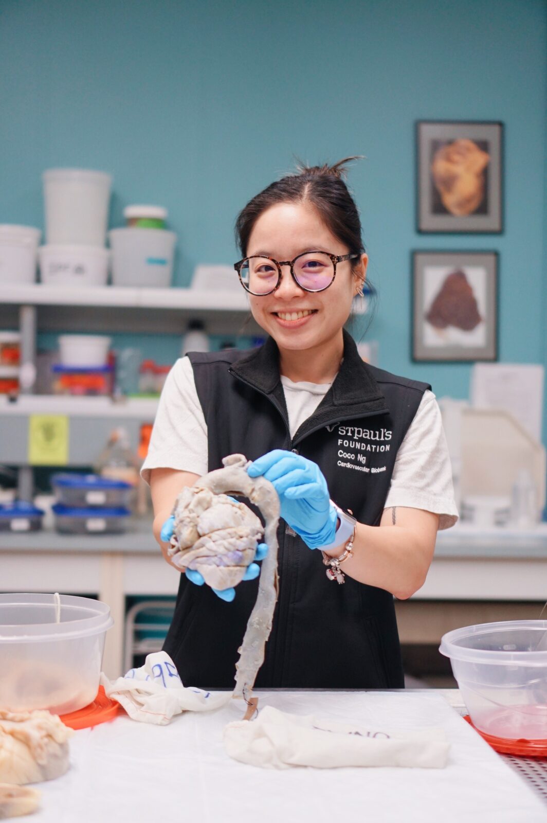
Have you ever wondered what happens to a heart removed (“explanted”) during a transplant surgery? You may think that because the heart is diseased, it would be of no use, discarded. But the journey of an explanted heart does not end upon removal. Each diseased heart still holds tremendous value for researchers. Heart researchers at the UBC Centre for Heart Lung Innovation, located in the heart of St. Paul’s Hospital, are dedicated to studying and understanding cardiovascular diseases. This includes giving explanted hearts a second home in our research laboratory, the BMCB. At the heart biobank, we are the final custodians of explanted hearts, preserving these patient-consented organs for research.
The BMCB is Western Canada’s largest and most comprehensive repository of human heart and cardiovascular tissue. Since 1982, the BMCB has collected over 100,000 specimens, including 550+ explanted hearts and other surgically removed tissues. These tissues are retrieved, anonymized, and meticulously processed, all within 24 hours! We use various preservation methods, such as flash-freezing and storing in long-term preservation chemicals, for a wide range of research studies. In a nutshell, this workflow is “biobanking” – collecting, processing, storing, and managing biological samples to advance scientific discovery.
The heart biobank provides researchers from around the world with high-quality tissue and clinically annotated patient data to accelerate discoveries in heart disease. As a biobank technician, I am responsible for all the heart tissue collected at the biobank. My role is critical in bridging the gaps between patients, clinicians, and researchers. I also help these groups understand how heart diseases develop and progress, connecting and mobilizing knowledge from bench to bedside.
2 Months Earlier – The Transplant
9:00PM – THE CALL
The alarm of my heart pager rings. This means that a heart transplant has been scheduled for the following day. It is typical for the heart biobank team to be paged at odd times of the day. Whenever heart transplants occur, multiple units, including the heart biobank team, BC Transplant Society, Cardiac Surgery Department, and Pre- and Post-Transplant Clinic, are involved to ensure the process is smooth and efficient.
St. Paul’s Hospital is British Columbia’s only site for adult heart transplants (BC Children’s Hospital performs pediatric heart transplants); therefore, all explanted hearts (from transplant) now reside with us, at the heart biobank. These hearts serve an important secondary purpose in supporting research, training, and education.
10:00AM – THE PREPARATION
Preparing for the retrieval of today’s explanted heart goes well. We get our tools and equipment ready and coordinate with the operating room team to ensure that the transplant is on schedule.
I go to meet today’s transplant recipient, Asher, to obtain informed consent for use of their explanted heart for research. They are hesitant and unsure about giving consent. I mention that regardless of their decision, they also have the opportunity to visit their explanted heart after surgery. All explanted hearts are kept at the biobank upon removal – even those that are not consented for research use.
Once these preparations are done, it is time for the waiting game.
1:00PM – THE BIOBANKING
The heart pager rings. The heart is out. I make my way to the operating room with purpose.
The heart biobank team has to act quickly to preserve the heart tissue before it starts to degrade. Once the explanted heart reaches the biobank, we swiftly begin our biobanking protocol. We first image, weigh, and note down any unusual observations. Then we begin sectioning out small pieces of the heart, including the greater arteries, valves, appendages, coronaries, and muscle. We preserve each of these sections of the heart by four different methods! These biobanking formats enable researchers to engage in a variety of research applications. Some are interested in examining genetic composition, microanatomy, and biomarkers that could lead to the development of therapeutics. Others are interested in how doctors can treat and prevent heart disease better with new tools and drugs. Even though we section out small pieces of the heart, after biobanking, the majority of the organ remains intact.
We follow a standard protocol when biobanking an explanted heart to ensure consistency and quality across each sample. It goes without saying that this standardization is important. What is truly remarkable is that each piece of the heart has the potential to lead to a discovery.
But every so often, the impact of these hearts extends far beyond the lab bench. Part 2, coming next week, follows one such moment: a reunion between a person and their own heart.
Coco Ng stands holding a fixed, human heart in the BMCB laboratory space. Photo by Tiffany Chang.