Services Cellular Imaging and Biophysics
Ultra high resolution imaging of cells and tissues
The Cellular Imaging & Biophysics Core at the Centre for Heart Lung Innovation provides services to individuals, institutions and industry in ultra high resolution imaging of cells and tissues in fixed and living specimens. Image analysis and consultations on microscopy related research projects are available upon request.
Confocal Microscopes
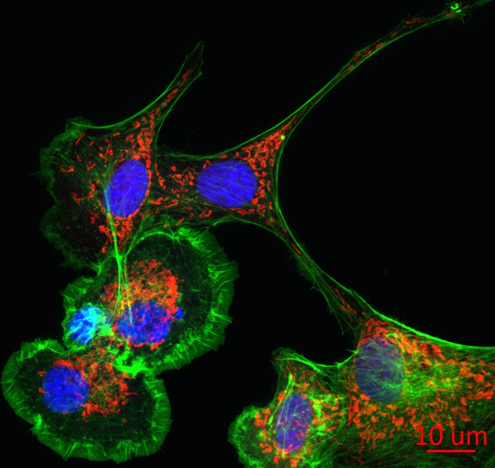
Our confocal microscopes can be used to 3-dimensionally image many biological systems and spatial distribution of fluorescently labeled proteins within a cell.
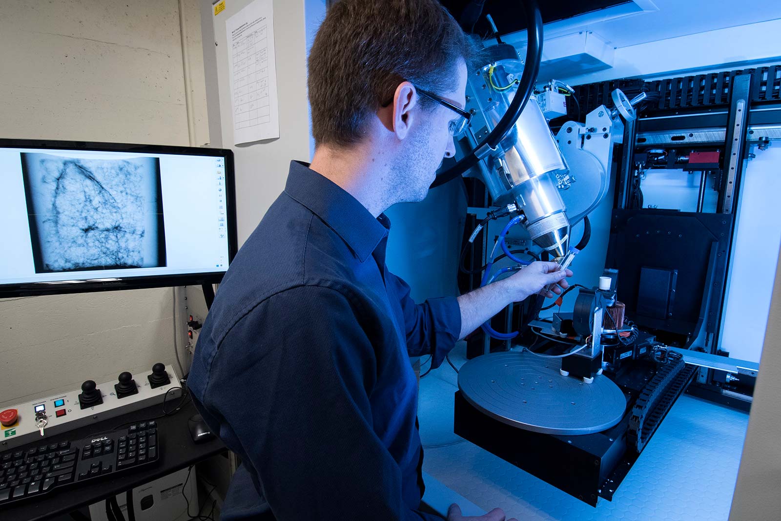
Micro Computed Tomography (micro CT) scanner
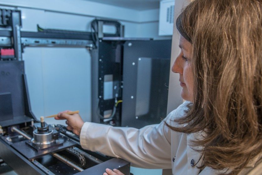
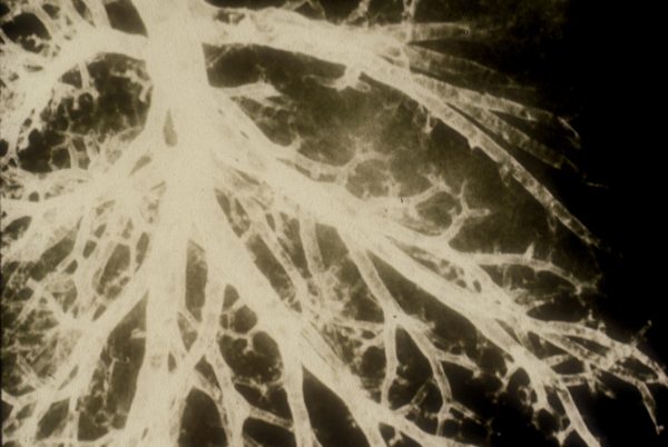
The Nikon micro CT scanner enables the micro-X-ray tomographic volume imaging of tissues and samples down to 1 micron, using the ultrahigh sensitivity PerkinElmer X-ray detector.
Wide Field Fluorescence Microscope
Wide Field Fluorescence Microscope and CCD camera which is capable of performing real-time, high speed imaging using an image splitter within the CCD camera that allows multi-wavelength fluorescence measurements to be taken in fixed and living cells.
Image Processing Stations
The Designated work stations are an excellent tool for 3D reconstruction and deconvolution of a series of 2D optical sections. The licensed Volocity software also enables the quantification and analysis of fluorescently labeled proteins or structures within an image.
Support
Our technicians have extensive training on all equipment and can provide support at every stage of your project from experimental design, protocol development, training and hands-on advice to data analysis.

Dragoş M. Vasilescu
PhD, Associate DirectorDr. Vasilescu, a Parker B. Francis Fellow, studies pulmonary diseases such as COPD and IPF using a combination of high resolution CT, histopathological microscopy and gene-expression tools.
He has taken on the role of Core director in 2014 and is leading the development of custom imaging protocols and image analysis methods for the PIs and students that use the core equipment.

Aaron M. Barlow
PhD, Core ManagerDr. Barlow is a specialist in optical and multiphoton microscopy as well as microCT imaging.
He uses his expertise to train, advise, and facilitate PIs and trainees in running imaging projects through the CIB.

