Services Magnetic Resonance Imaging
Advanced Imaging of Patients with Heart and Lung Disease
The Magnetic Resonance Imaging (MRI) Core at the Centre for Heart Lung Innovation provides access to state of-the-art MRI equipment and protocols dedicated to structure-function investigations of heart and lung disease.
3T MRI
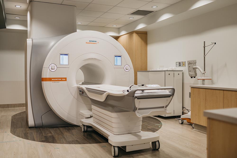
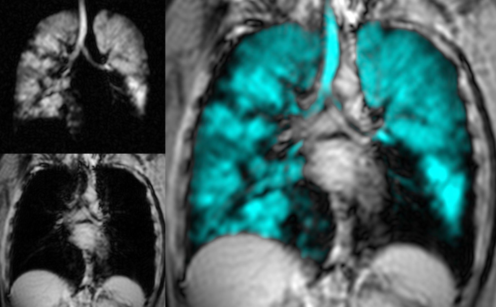
Central to the facility is a Siemens Vida 3.0 Tesla clinical MRI scanner with a 70 cm bore and full complement of anatomy-specific RF coils for human imaging. This scanner includes broadband RF hardware capable of imaging hyperpolarized 129Xe gas as an inhaled contrast agent. Advanced research protocols are available for structure-function cardiac and pulmonary imaging.
Hyperpolarized 129Xe Gas Facility
The Polarean 129Xe Hyperpolarizer enables high-efficiency polarization of 129Xe gas as a contrast agent in combination with dedicated 129Xe lung RF coils for advanced functional lung imaging.
Support
Our clinical and research technologists have extensive training on all MRI and polarizer equipment. Support is available at every project stage including experimental design, 129Xe MRI and other imaging-related REB applications, protocol development, training and hands-on advice for image and data analysis.

Rachel Eddy
MRI Core DirectorDr. Rachel Eddy is an imaging scientist with expertise in quantitative CT and MRI of the lungs.
Dr. Eddy launched the pulmonary MRI research program using hyperpolarized 129Xe gas at HLI. Her research is focused on developing and applying novel pulmonary imaging tools to better understand pathophysiology of lung disease. As the MRI Core Director, Dr. Eddy leads the development of MRI protocols and image analysis methods, directing the 129Xe Hyperpolarized Gas Facility.

